Electron diffraction: A tool for hydrogen atom localization and absolute structure determination of nanocrystals containing organic molecules
Petr Brázda¹*, Lukáš Palatinus¹
¹ Institute of Physics of the Czech Academy of Science, 182 21 Prague, Czech Republic
* Corresponding author: brazda@fzu.cz
Organic molecular crystals and metal organic frameworks (MOFs) are two families of materials, which often only form submicrometric crystals, whose structure cannot be characterized by conventional single-crystal x-ray diffraction techniques. During the last decade, 3D electron diffraction (3D ED) developed to a method fully suitable for the structure solution of nanoscale materials.
Organic molecular crystals and MOFs pose specific problems for the structure analysis by 3D ED. They often exhibit rather large mosaicity, they tend to contain disorder and on top of this they are very beam sensitive. Therefore, the determination of crystal structures of these materials is a challenging task. The problem of radiation damage may be mitigated by sample cooling, low dose techniques, distribution of the dose to a larger volume through crystal scanning in the case of rod-shaped or platelet crystals or combination of datasets from more crystals. In the most challenging cases, several or all of these approaches need to be combined. An important recent development has also been the advent of direct detection cameras, which enables acquisition of essentially noise free data. Without all these developments it would not be possible to solve a large portion of interesting materials. In our group, we develop software PETS [2], which in its current version 2.0 provides a complete data processing work flow and contains several advanced features specific for 3D ED data like treatment of precession electron diffraction data, frame orientation refinement and correction for optical distortions. These advanced tools are vital for structure solution of beam sensitive materials as they significantly improve the quality of intensity extraction from the acquired data.
Dynamical refinement (application of dynamical diffraction theory), which is developed in our group allowed more robust determination of the fine structural details like localization of the hydrogen atoms [3] and it enabled determination of the absolute structure of a chiral material [4]. These features are of eminent interest for example for pharmaceutical industry. About 90% of the active pharmaceutical ingredients (APIs) are chiral molecules and determination of the absolute configuration is necessary for the drug approval. Determination of the position of protons, i.e. the ability to distinguish between salt and cocrystal, is also an important problem with potential impact on the patent protection of the drugs. Understanding the functionality of MOFs sometimes also requires the localization of hydrogen atoms as potential sites of catalytic activity.
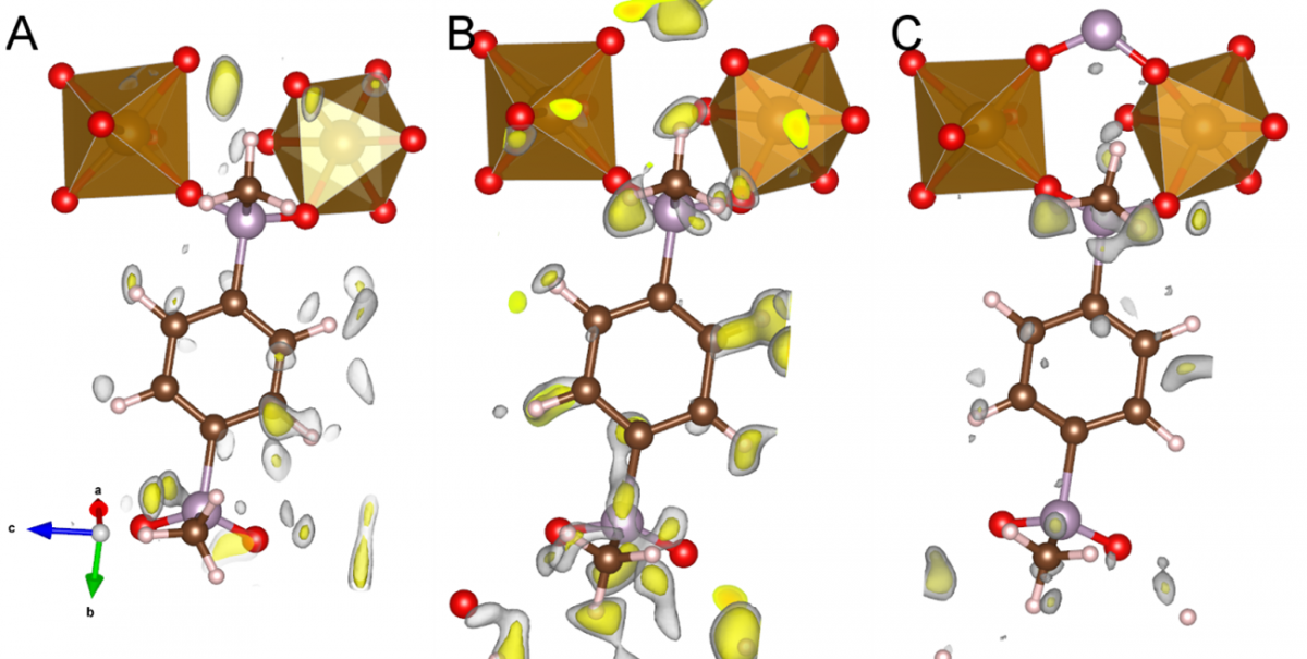
Figure 1 Difference potential maps at 2.0 sigma (grey) and 2.5 sigma (yellow) of ICR-2. Dynamically refined data processed without frame orientation refinement (A), dynamically refined data processed with frame orientation refinement (B), kinematically refined data processed with frame orientation refinement (C).
[1] M Gemmi et al., ACS. Central. Sci. 5 (2019) p. 1315.
[2] L Palatinus et al., Acta Crystallogr. B75 (2019) p. 512.
[3] L Palatinus et al., Science 355 (2017) p. 166.
[4] P Brázda, L Palatinus and M. Babor, Science 364 (2019) p. 667.
[5] This research was supported by the Czech Science Foundation, project number 20-04408S. [6] We acknowledge CzechNanoLab Research Infrastructure supported by MEYS CR (LM2018110).
[7] The authors thank Zentiva k.s. and The Parc (https://www.theparc.eu/) for providing the samples.